


Reducing Occupational Exposure During PET-CT Scans with an Automated Injection System
KATE VAN ELTEREN[1], IAN HUFTON[1], DANIEL CARVALHO[1]
BNMS (British Nuclear Medicine Society) Spring 2024– Belfast (Irlande du Nord) – May 13-15, 2024
[1] Royal Liverpool Hospital, Mt Vernon Street, Liverpool, L7 8YE
Occupational radiation doses received by staff performing PET-CT scans are generally among the highest recorded in nuclear medicine staff (e.g., Skovorodko et al., 2020).
This elevated exposure is due to several factors:
- the use of a positron emitter as a diagnostic imaging agent (primarily 18F at the Royal Liverpool University Hospital (RLUH));
- the high-energy annihilation photons produced, which are challenging to shield effectively;
- the large number of patients handled daily (approximately 20 patients/day at RLUH);
- and the additional clinical demands of many patients due to co-existing conditions that are often more complex than those seen in traditional nuclear medicine.
Injecting patients and preparing individual doses for injection from multi-dose vials contribute significantly to the overall occupational dose of staff performing PET examinations. Together, these two activities account for over 40% of the annual whole-body dose, with 30% attributed to injections (Peet et al., 2012).
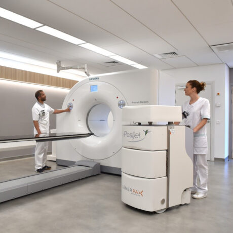
Automated injection systems such as Posijet® (Lemer Pax) offer advantages like reducing human error during dose preparation, and can also reduce sources of occupational exposure in several ways:
- By minimising operator contact with the vials by isolating the stock solution in a shielded environment and automatically diluting/preparing doses inside the unit.
- By eliminating the need to handle syringes thanks to administration kits that directly connect the unit to the patient.
- By providing extra protection for operators during patient injections.
- By reducing the risk of needlesticks and spills during manual preparation and injection.
METHOD:
To assess the benefits of the Posijet® system (introduced in 2022 at RLUH for PET-CT staff), an audit of radiation doses (Landauer; whole-body, finger, and eye) was conducted by the medical physics team before and after the system’s implementation.
Data spanning 47 months prior to Posijet® implementation and 13 months post-implementation were collected. Ring and dosimeter data were analysed for two staff members who continuously performed PET-CT scans during both periods, with no extended absences and similar work schedules. Eye dosimeter data were analysed for one staff member.
RESULTS:
The audit results are presented in the table and graphs. To aid interpretation, dosimeter results are presented as monthly averages, with the graphs showing annual averages as a time series, along with a pre-Posijet overall average for context.
To give an idea of the variability, standard errors of the mean (SEM) are reported for the annual mean of each participant, and t-tests were used to assess significance (p < 0.05). A correction factor was applied to the compiled annual averages to account for the number of patients.
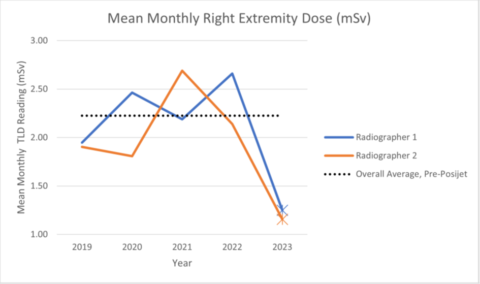
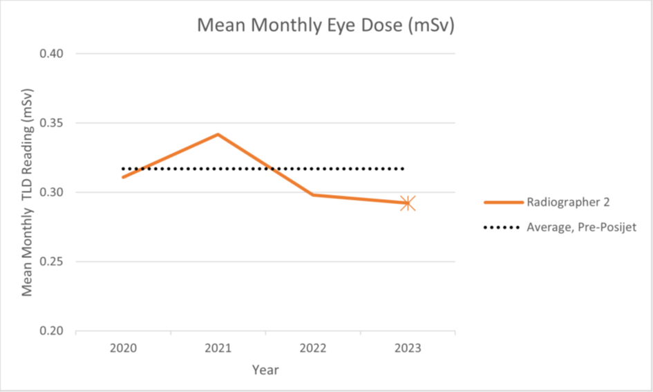
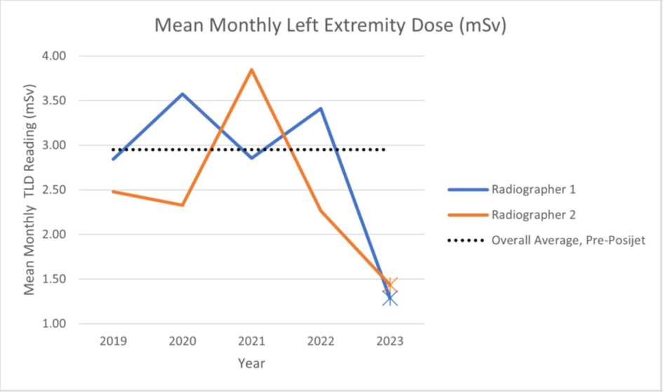
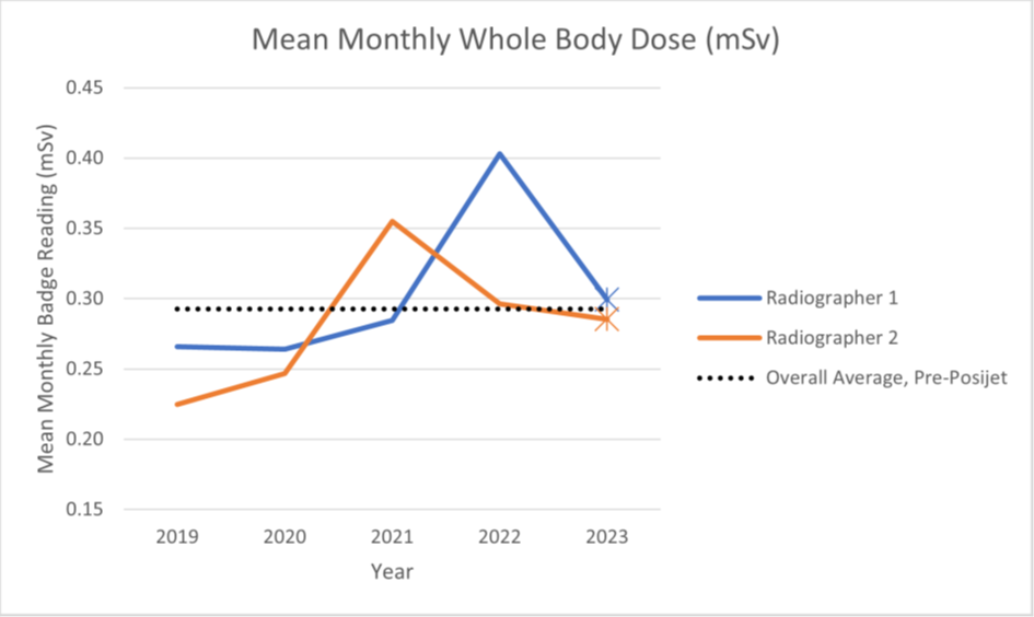
GRAPHS: Average monthly occupational doses for the two radiographers working continuously in the PET-CT department during the entire data collection period. A sharp reduction in the extremity dose and a slight reduction in eye dose were observed, with no definitive change in whole-body dose.
For the participating radiographers whose exposure data we analysed, the following trends were observed:
- Extremity dose reduction: Over 100% (117% for the left hand; 85% for the right hand; SEM 7%–25%; p < 0.001).
- Eye dose reduction: 9% (SEM 12%–21%; p > 0.1).
- No clear change in whole-body dose: <1% (SEM 6%–17%; p > 0.1).
The Posijet®’s benefit in reducing extremity exposure is clear; there is a striking contrast between pre- and post-Posijet® extremity dosimeter (ring) results. This aligns with findings by Covens et al. (2010), who reported a 95% reduction in extremity doses.
| Participant | Whole-body dose reduction (%) | Left-extremity dose reduction (%) | Right extremity dose reduction (%) | Eye dose reduction (%) |
| Radiographer 1 | 1.7 [46,9] | 146.3 [43,9] | 85.2 [42,8] | |
| Radiographer 2 | -1.6 [44,9] | 90.1 [43,11] | 84.7 [43,11] | 8.5 [20,9] |
| TOTAL | 0.1 | 116.7 | 84.9 | 8.5 |
DISCUSSION:
Since its clinical introduction, the Posijet® system has greatly reduced extremity doses for staff performing PET-CT scans by minimising the need to handle and inject unsealed radioactive sources. Additionally, the reduced handling of sharp radioactive objects lowers the risk of needle stick injuries, which could otherwise exceed the annual professional skin dose limit of 500 mSv under the Ionising Radiations Regulations (IRR) when using 18F, the main PET imaging isotope used at RLUH (calculated locally using Varskin+).
While we found no reduction in whole-body dose or strong evidence of eye dose reduction, these findings are limited by the small number of participants. Moreover, injection is not the sole contributor to whole-body dose. Covens et al. (2010) reported a 20% reduction in whole-body dose with their system, attributing this comparatively modest reduction to the fact that patient positioning contributes approximately four times more to whole-body dose than injection (slightly lower than Peet et al.’s 2012 estimates).
Nevertheless, our findings prompted a wider audit of the doses received by staff performing PET-CT scans using an EPD dosimeter to track and correlate accumulated radiation exposure with specific professional activities. This aimed to inform best practices and included a follow-up audit of staff dosimeters in the subsequent calendar year to gather additional data.
Although the Posijet® has demonstrated its potential for reducing extremity doses in RLUH staff and performed well in meeting the department’s needs, several factors should be taken into account when considering the acquisition of such a system:
- Training: Understanding system operation, troubleshooting, etc.
- Quality Assurance/Maintenance: integrated dose calibrator, system connectivity, injection kits
- Frequency of multi-dose injection procedures versus single-dose injection procedures
- Overhead Costs: vial kits (one per vial) and individual patient kits (one per patient).
- The number of patients in a given calendar year
- Timing between procedures: initial system set-up at RLUH takes 20-30 minutes for experienced users, and vial changeover takes 15-20 minutes
- The suitability of other available PPE: automated benchtop dose preparers, vial shields/syringe shields.
The authors declare no conflicts of interest related to this presentation.